Cell Organelles and Reproduction
|
|||
|
Major Cellular Organelles |
Function |
Prokaryotic |
Eukaryotic |
|
Cell Membrane |
Selective Barrier Transport of cellular nutrients and waste Recognition of cellular signals |
Y |
Y |
|
Cytosol |
Interior matrix of cell Contains cellular molecules, macromolecules such as enzymes, lipids, carbohydrates, and RNA Organized and compartmentalized |
Y |
Y |
|
Nucleus |
Largest organelle Contains chromosomes Chromosomes consist of DNA and Protein Organized by protein matrix |
N |
Y |
|
Mitochondria |
Contains the Kreb cycle and electron transport system Major site of ATP synthesis Involved with cellular “self destruct” called Apoptosis |
N |
Y |
|
Chloroplast |
Contains the Photosynthetic systems Responsible for converting light energy to chemical energy |
N |
Y |
|
Ribosome |
Composed of RNA and proteins Responsible for synthesizing protein molecules http://micro.magnet.fsu.edu/cells/animals/images/ribosome.jpg |
Y |
Y |
|
Lysosome |
Contains digestive enzymes that break up proteins, lipids, and nucleic acids Responsible for removing waste molecules and recycling molecular subunits http://micro.magnet.fsu.edu/cells/animals/images/lysosome.jpg |
N |
Y |
|
Consider how some
of these organelles interact: GENE
EXPRESSION: nucleus- (DNA > gene > transcription
produces m RNA > mRNA leaves through nuclear pore into cytosol> binds to ribosome and directs
the synthesis of a protein. CELL MEMBRANE : contains proteins that bind molecules > after
binding, protein is endocytosed (http://www.sp.uconn.edu/~bi107vc/images/cell/clathrin.jpg)
> protein releases the molecule and can be reused by exocytosis
(reverse of endocytosis) or can be sent to lysozyme for degradation CELL
REPRODUCTON: many organelles are
involved in cell reproduction, including the nucleus and the mitochondria and
chloroplasts. All of these organelles
contain DNA that must be replicated so that each new cell will have complete
copies. Prokaryotic cells
(bacteria) divide by: 1.
Duplicating
their circular DNA molecule 2.
Attaching each
DNA molecule to the cell membrane 3.
Adding membrane
components between the attachments to separate the duplicated DNA molecules 4.
Using membrane
fission (http://bio.winona.msus.edu/bates/genbio/images/fission.gif)
to create two new bacteria cells Eukaryotic cells
divide by: 1.
Duplicating the
chromosomes (chromosomes contain DNA and protein) 2.
Separating the
duplicated chromosomes (Mitosis) 3.
Duplicating
other organelles (mitochondria, chloroplast, etc) 4.
Reforming the
nuclear envelope 5.
Laying down
membrane components between the two nuclei (cytokinesis) 6.
Forming two new
cells (http://genetics.gsk.com/graphics/mitosis-big.gif)
|
||||||
ACTIVITY-
1) Compare
the rate of reproduction between a bacteria (simple asexual)
and a eukaryotic cell (complex asexual) using www.cellsalive.com . Students use graph paper to plot the growth
curve and compare the two types of cells. The web site Cells Alive.com has many
resources for studying cells and cell structure. The resource that we are going to utilize
is the Cell A conceptual method for teaching
students the difference between prokaryotic and eukaryotic cells is for the
students to answer the question: Which cell type reproduces faster? This
question can be best answered by comparing the growth rates of the two
different cell types. Since it is
difficult and costly to set up cell cultures, we will utilize the data from
the Cell Collecting the data from Cell Bacterial Cell Cam
(note- the images were collected every 5 minutes) Cancer Cell Cam (note- the images
were collected every 10 minutes) Items needed for activity: ·
Graph paper ·
Images from the cell cam. 2) Examine
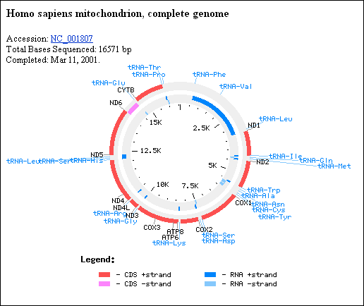
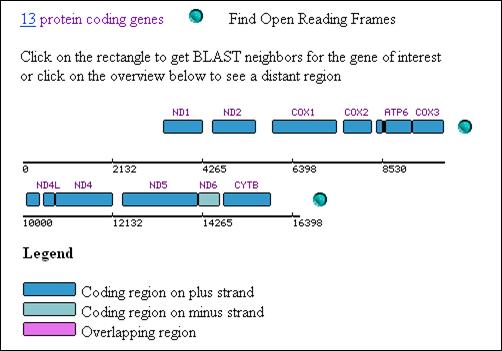
maps of Mitochondrial, Chloroplast, and Genomic DNA using online resources (http://www.ncbi.nlm.nih.gov/) To demonstrate the use of web based
resources concerning DNA and biotechnology, we will examine using the NCBI ( http://www.ncbi.nlm.nih.gov/). We will compare the sizes and complexity of
the mitochondrial and chloroplast genomes as an example of the types of
information one can gather at this site. ·
First we can examine a map of the human
mitochondrial DNA at http://www.ncbi.nlm.nih.gov/genomes/framik.cgi?db=Genome&gi=12188
·
You can
use this type of image to show the students the minimum genes that are found
on the human mitochondria. Also, you
can point out that for many of the mitochondrial proteins, other genes are
located in the nucleus; thus it requires both sets of genomes to make
functional mitochondria. For example:
|